New test

About retinal vein occlusion (RVO)
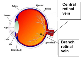
A retinal vein occlusion (blockage) occurs when one of the retinal veins of the eye becomes blocked. (The retina is the light sensitive layer of the back of your eye).
There are two types of vein occlusion, depending on which retinal vein is blocked:
- Branch retinal vein occlusion (BRVO). This blockage involves one of the four retinal veins. Each one of these veins drains the blood of a quarter of the retina.
- Central retinal vein occlusion (CRVO). This blockage occurs in the main vein of the retina. The loss of vision is generally more severe.
Retinal vein occlusion and effect on vision
Retinal vein occlusions can cause different complications in the eye. These include the following.
Macular oedema: This is the collection of fluid or swelling in the back of the eye, affecting the central vision. There are several treatment options for this.
Ischaemia: This is damage to the retina because of the obstructed blood flow and lack of oxygen. There is no treatment available for this.
Neovascularisation: This is abnormal blood vessel growth, either in the retinal surface or in the front of the eye (iris). This can lead to bleeds and/or increased eye pressure. Treatment is available for this.
What causes a retinal vein occlusion?
The exact reason for the blockage in the retinal veins is generally unknown, but certain conditions can make it more likely to occur. These include:
- high blood pressure
- diabetes
- high cholesterol
- smoking
- certain blood disorders
- glaucoma.
It is important that we carry out tests to detect risk factors and minimise the risk of you developing further vein occlusions, either in the affected eye or in the other eye. Further vein occlusions affect 5 to 10% of patients.
Testing for RVO
Your doctor may recommend that we carry out the following tests.
Optical Coherence Tomography (OCT)
Test for macular oedema
Procedure: This is a non invasive retinal scan that allows us to make an exact measurement of the swelling in the back of your eye.
Fundus fluorescein angiography (FFA)
Test for Ischaemia and Neovascularisation
Procedure: We can do this because the dye circulates through your retinal blood vessels, allowing us to make an accurate assessment of the retinal blood supply (ischaemia) and the possible development of abnormal blood vessels (neovascularisation).
You may also need a blood test to investigate possible risk factors (see table below).
Treatments for RVO, information and risks
Macula oedema (persistent swelling in the back of the eye) is the main cause of vision loss in patients with retinal vein occlusions.
Intraocular injections of antiVEGF (Eylea, Lucentis, Ongavia, Vabysmo).
This is currently the first treatment option for macular oedema.
These injections are given every month, at the beginning of the treatment, and then at extended intervals, depending on your response. They are given with local anaesthetic.
More than 50% patients achieve a significant gain of vision with this treatment (improvement of 3 lines in a standard vision chart). There is a 1 in 1000 chance of vision loss as a complication of these injections.
Let your doctor know if you are pregnant, have an infection or had a recent heart attack or stroke. In these situations, these injections may not be a suitable treatment for you.
Intraocular implants of steroids (Ozurdex)
These can be given every 4 to 6 months for macular oedema. They are given with local anaesthetic.
More than 50% patients will achieve a significant gain of vision with this treatment (improvement of 3 lines in a standard vision chart). There is a 1 in 1000 chance of vision loss as a complication of these implants.
Note that these implants can accelerate the formation of cataracts and increase the pressure of the eye, which can result in glaucoma.
Laser photocoagulation
In some cases this may be advised by your doctor, especially if you have developed abnormal blood vessels (neovascularisation) or are at high risk of developing them.
Observation
This is sometimes considered.
Emergency help, hours of operation and referral system
Kingston Hospital Eye Casualty, Galsworthy Road, KT2 7QB
8.30am to 4.30 pm (last appointment at 4pm).
Closed on weekends and bank holidays.
Booked appointments only. Call to book on 020 8934 6799.
Western Eye Hospital Emergency Department, 153 Marylebone Rd, London NW1 5QH
8am to 8.30pm, every day.
Walk in service, no referral required.
Moorfields Eye Hospital, 162 City Road, London EC1V 2PD
24 hours a day, every day.
Walk in service, no referral required.
020 7566 2345 or 020 7253 3411.
Moorfields Eye Clinic at St George’s Hospital,Tooting, London SW17 0QT
4.30pm to 8.30pm on weekdays, 24 hours on weekends.
Referral via an emergency GP or booked appointment required.
Call to book on 020 7702 5542.
Contact information
Kingston Hospital Royal Eye Unit
Kingston Hospital
Galsworthy Road
Kingston KT2 7QB